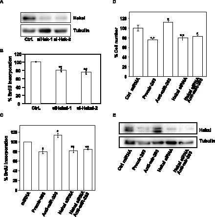
Figure 5. Influence of miR-203-regulated Hakai on cell proliferation.

A, levels of Hakai and loading control α-tubulin were tested in whole-cell lysates by Western blotting 48 h after transfecting HeLa cells with two different siRNAs oligos for Hakai relative to the scrambled Ctrl small RNA group. B, 48 h after transfection of HeLa cells with the indicated siRNA oligos, cell numbers were measured by BrdU incorporation assay and represented as percentage of cells. C, measurement of BrdU incorporation 48 h after transfection of HeLa cells with the indicated small RNAs. D, measurement of cell number by hemocytometer after 48 h of transfection of HeLa cells with the indicated small RNAs. E, forty-eight h after transfecting HeLa cells with the indicated small RNAs, Hakai and α-tubulin levels were measured by Western blot analysis. Western blotting data are representative of three independent experiments. The data in panels B–D are the means ± SEM from three experiments. Significant differences in BrdU incorporation (B and C) and in cell number (D), compared to the transfected scrambled (Ctrl) small RNA are indicated (*p<0.05, **p<0.01).
