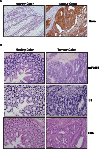
Figure 6. Hakai and miR-203 expression in colon tissues.

A, immunohistochemical analysis of Hakai protein expression in normal colon versus colon cancer tissues. Differences between tumour samples compared to its paired healthy tissues are statistically significant (***p<0.001, n = 19). B, in situ hybridization (ISH) for miR-203 (upper panel) and U6 snRNA (as control probe, middle panel) expression in colon cancer tissues compared to normal colon tissue. Haematoxylin and eosin (H&E)-stained section (lower panel) was included to identify tumour area. Scale bar, 200 µm.
