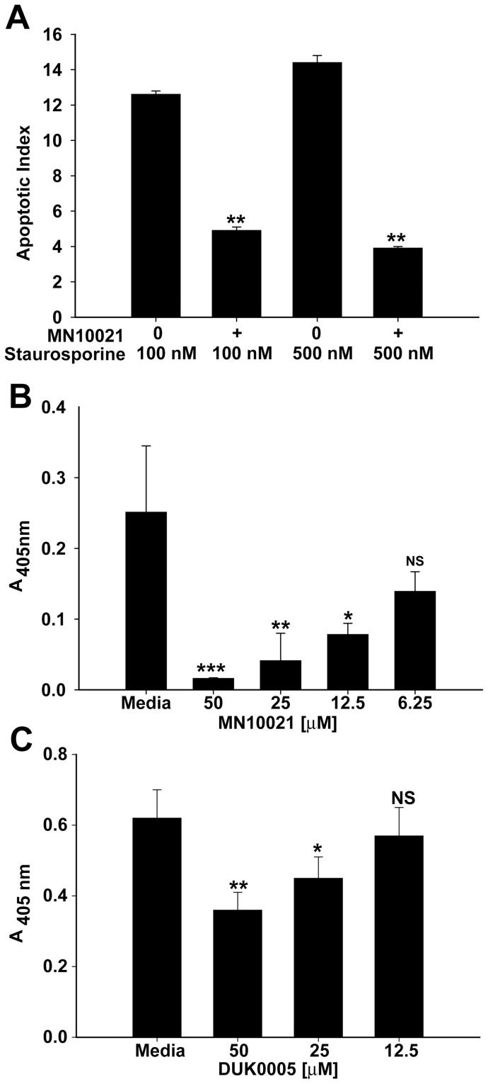
Figure 10. MN10021 and DUK0005 inhibit apoptosis and necrosis in HUVEC.
A. HUVEC were grown to confluence in 96-well tissue culture plates. Media was removed from the wells and to each of quadruplicate wells was added 200 µl of either fresh media (untreated) or fresh media containing 50 µM MN10021 (treated) and the cells incubated 3 h at 37°C. To quadruplicate wells of both untreated and treated cells were added 20 µl of media or 20 µl of media containing either 100 or 500 nM staurosporine. The plates were incubated overnight at 37°C and the cells processed for measurement of apoptosis using the Cell Death Detection assay (Roche Diagnostics) per the manufacturer's instructions. The apoptotic index was calculated as described in Methods. B. HUVEC were grown to confluence in 96-well tissue culture plates. Media was removed from the wells and to each of quadruplicate wells was added 200 µl of either fresh media (untreated) or fresh media containing 50, 25, 12.5, or 6.25 µM MN10021 (treated) and the cells incubated 3 h at 37°C. To quadruplicate wells of both untreated and treated cells were added 20 µl of media containing 100 ng/ml human TNF-α (recombinant; R & D Systems). The plates were incubated overnight at 37°C and the supernatants removed and processed for measurement of necrosis using the Cell Death Detection assay (Roche Diagnostics) per the manufacturer's instructions. Absorbance values for the supernatants of untreated cells (no TNF-α) were negligible. C. HUVEC were grown to confluence in 96-well tissue culture plates. Media was removed from the wells and to each of quadruplicate wells was added 200 µl of either fresh media (untreated) or fresh media containing 50, 25, 12.5 µM DUK0005 (treated) and the cells incubated 3 h at 37°C. To quadruplicate wells of both untreated and treated cells were added 20 µl of media or 20 µl of media containing 100 nM staurosporine. The plates were incubated overnight at 37°C and the cells processed for measurement of apoptosis using the Cell Death Detection assay (Roche Diagnostics) per the manufacturer's instructions. Absorbance values for untreated cells (no staurosporine) were negligible. *** p<0.001; ** p<0.01; * p<0.05.

