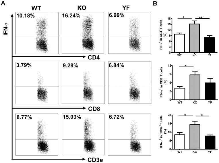
Figure 4. IFN-γ producing T cells in the lung increased in ICOS-KO mice but remained the same in ICOS-YF mice.
Lung mononuclear cells were prepared at day 14 post-infection and the IFN-γ producing cells were quantified by intracellular cytokine staining as described in Materials and Methods. Representative flow cytometric plots (A) and a summary of the results (B) are shown. Data are presented as mean ± SD (7 ICOS-WT, 4 ICOS-KO, and 7 ICOS-YF mice) from one representative experiment out of three independent experiments with consistent results. * p<0.05, ** p<0.01.

