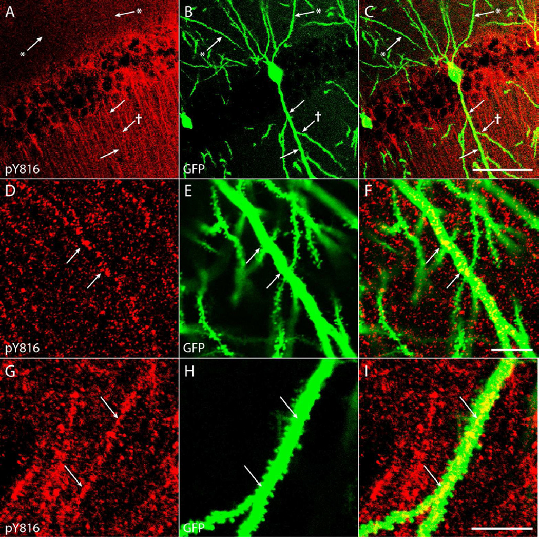
Figure 7. pY816 TrkB immunoreactivity is intracellular and punctate within the dendritic shaft of proximal apical dendrites of CA1 pyramidal cells.
A–C) The primary apical dendrite of a GFP+ CA1 pyramidal neuron (B; arrows) contains clear pY816 immunoreactivity (A; arrows) within its aspinous proximal shaft, whereas a secondary dendritic branch (B; arrow with dagger) does not contain prominent immunoreactivity within its shaft (A; arrow with dagger). Basal dendritic processes (B; arrows with asterisks) clearly do not contain pY816 immunoreactivity (A; arrows with asterisks). Merged pY816 and GFP is shown in (C). Image is a maximum projection (displaying the region of highest intensity) of multiple confocal scans taken at 0.4 µm increments in the z-plane. Scale bar = 50 µm. D–F) Close-up of a proximal, aspinous portion of a GFP+ apical dendritic process (E; arrows) with intracellular, punctate pY816 immunoreactivity (D; arrows) within the shaft. Merged pY816 and GFP is shown in (F). Scale bar = 10 µm. G–I) A spiny portion of a GFP+ apical dendrite (H; arrows) also displaying intracellular, punctate pY816 immunoreactivity (G; arrows) within the shaft. Merged pY816 and GFP is shown in (I). Scale bar = 10 µm.

