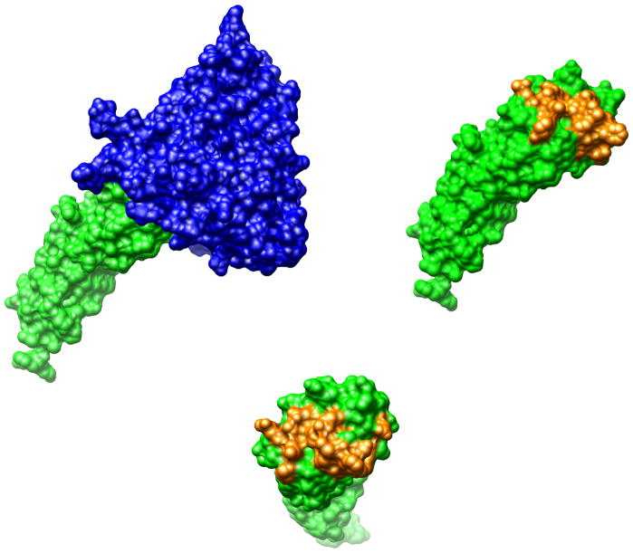
Figure 2.
The structure of gp120 (blue) in complex with CD4 (green) extracted from pdb file 1G9N. The protein/protein interaction surface extends to about 2,000 Å2. The residues in CD4 that comprise most of the interaction with gp120 (Gln 25, His 27, Lys 29, Gln 33, Lys 35, Asn 39, Gln 40, Ser 42, Phe 43, Leu 44, Thr 45, Pro 48, Arg 59, Asp 63 and Glu 85) are shown in yellow. Single amino acid sCD4 mutants in which these amino acids were individually mutated to alanine were used in the analysis presented in this paper.

