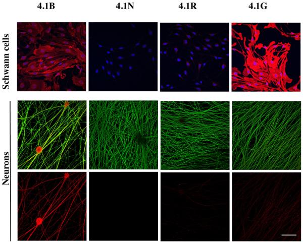
Figure 1. Expression of 4.1 proteins by neurons and Schwann cells.
Immunostaining of Schwann cell and DRG neuron cultures for different members of the 4.1 family is shown. Schwann cells (top row) were double stained with Hoechst (blue) to demonstrate cell nuclei and for 4.1 proteins (red). Neurons (middle and bottom rows) were double stained for neurofilament (green) and 4.1 proteins (red) in the middle row and 4.1 proteins only, in the bottom row. Schwann cells express 4.1B at moderate levels and 4.1G at robust levels; neurons principally express 4.1B. Scale bars: panels A and B, 5 μm; panels C-F, 0.5 μm; bar 50 μm.

