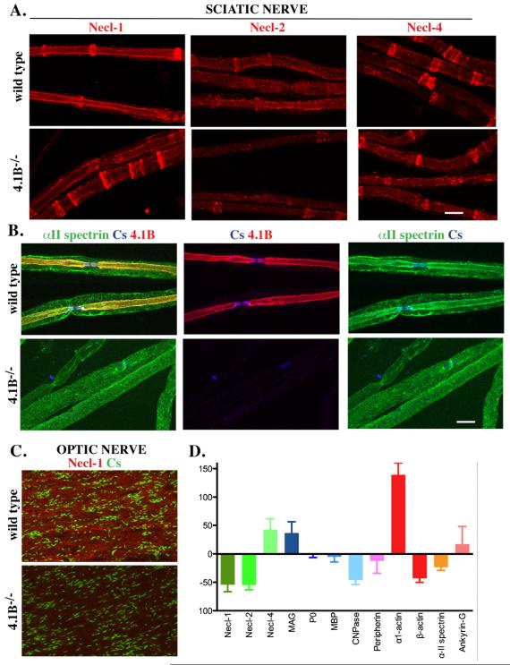
Figure 5. The expression of internodal components are altered in 4.1B−/− nerves.
A. Teased sciatic nerves from wild type and 4.1B−/− mice were stained for the Nectin-like proteins. There is a modest reduction of Necl-1 and Necl-2 along the internode but not the clefts; Necl-4 levels are largely unchanged. B. α2 spectrin staining co-localized with 4.1B along the internode of sciatic nerve fibers in wild type mice (yellow in the merged image). The inner staining that colocalizes with 4.1B along the axon was markedly reduced in the 4.1B−/− nerves. C. A reduction of Necl-1 staining was observed in the optic nerve of the 4.1B−/− mice although individual axons were not readily visualized. D. Summary of Western blotting data analyzing expression of myelin proteins, domain components and cytoskeletal proteins from three sets of sciatic nerves; the levels of these proteins in 4.1B−/− mice are shown normalized to their levels in wild type mice. Scale bars (A, B) 10 μm.

