
FIGURE 1. Initial and Collecting Lymphatics.
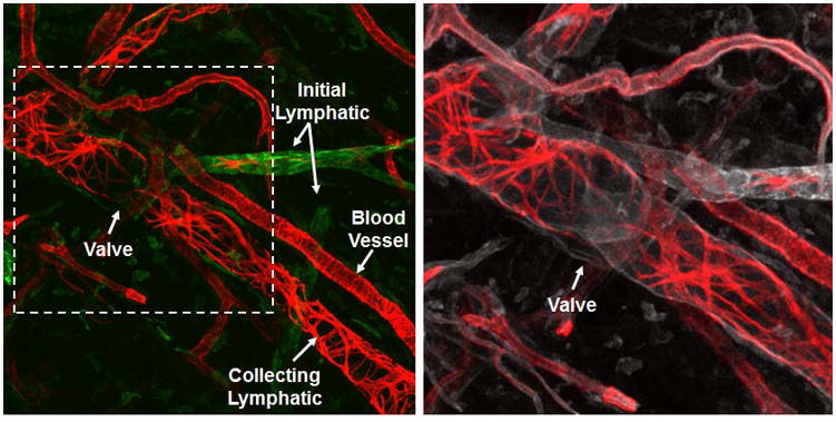
The lymphatic vessels of the ear of an athymic nude mouse are shown. LYVE-1 (green) indicates the initial lymphatic vessels. αSMA (red) indicates the SMCs of the collecting lymphatic vessels and blood vessels. The circumferential αSMA staining pattern of the collecting lymphatic vessels is distinct from the more homogenous pattern of the blood vessels. CD31 (white) indicates all endothelial cells in the field and shows an intraluminal valve in the collecting lymphatic vessel.