
Figure 1.
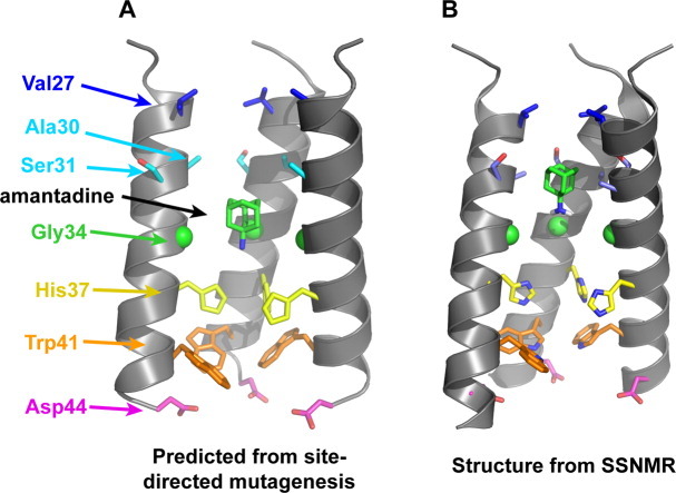
(A) Functional model of the TM domain of the M2 tetramer, showing the positions of crucial side chains and the drug amantadine. The model was obtained from cysteine scanning mutagenesis.3,4 (B) High-resolution structure of the amantadine-bound M2TM in DMPC bilayers obtained from SSNMR (PDB: 2KQT).5 The overall shape of the tetrameric bundle from the functional model is in excellent agreement with the high-resolution structure; however, specific differences exist such as the helix tilt angle and the conformations of several side chains (e.g., Ser31 and Trp41). In both images, the “front” helix has been removed for clarity.