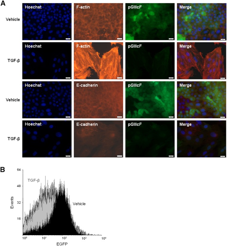
FIGURE 3.
Fluorescence-based splicing reporters mark epithelial–mesenchymal transitions (EMT) in vitro. (A) Normal mouse mammary (NMuMG) cells stably transfected with pGIIIcI2 were cultured in the presence of TGF-β or vehicle for 7 d. Addition of TGF-β resulted in a marked decrease in EGFP expression (second column of images), and the loss of EGFP was related to the formation of actin stress fibers (third column, top two rows) and down-regulation of E-cadherin (third column, bottom two rows). (B) NMuMGs reduced EGFP expression by nearly threefold after incubation with TGF-β, as estimated by flow cytometry.

