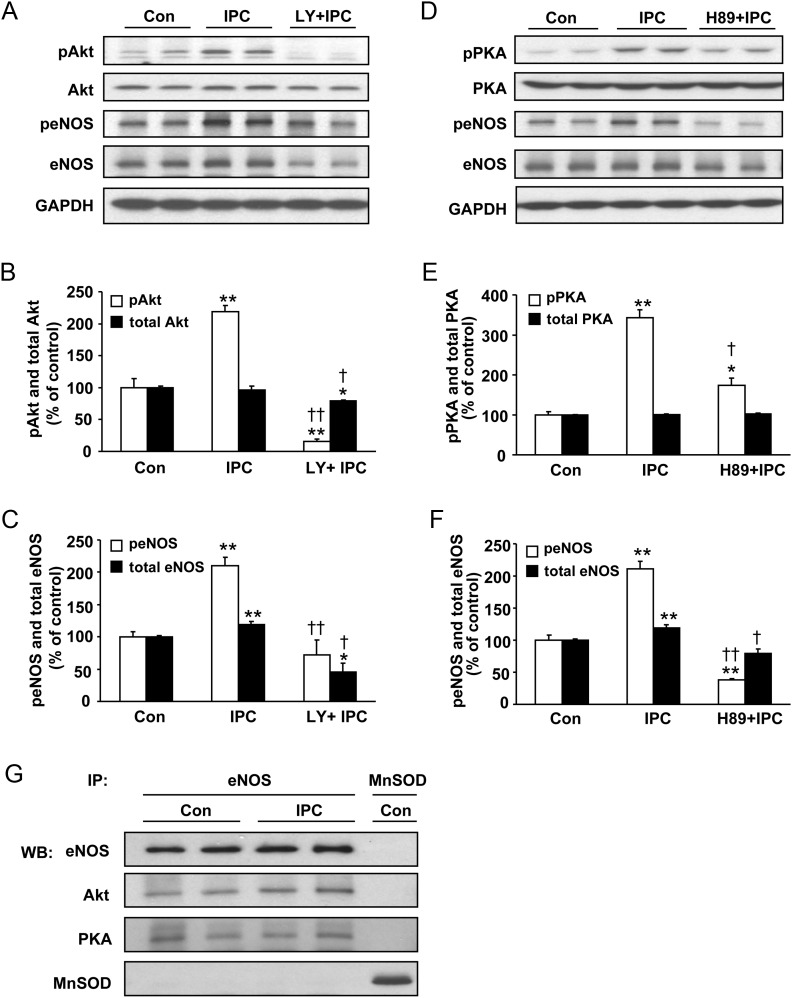
Figure 4.
The effect of inhibition of Akt (A–C) or PKA (D–F) on eNOS phosphorylation following IPC induction; and association of Akt and PKA with eNOS in control and IPC hearts (G). Representative immunoblots of hearts subjected to control perfusion (Con), IPC and IPC + PI3K/Akt inhibitor LY (15 µM) for pAktSer473 (pAkt), Akt, peNOSSer1176 (peNOS) and eNOS (A), with quantitative bar graphs of pAkt and peNOS in (B) and (C). Representative immunoblots of hearts subjected to Con, IPC, and IPC + PKA inhibitor H89 (10 µM) for pPKAThr197 (pPKA), PKA, peNOSSer1176 (peNOS), and eNOS (D), with quantitative bar graphs of pPKA and peNOS in (E) and (F). Values are means ± SE. n = 4/group. *P < 0.05, **P < 0.01 vs. control hearts. †P < 0.05, ††P < 0.01 vs. IPC alone. (G) Immunoprecipitation (IP) of eNOS resulted in co-precipitation of Akt and PKA in heart tissues subjected to control perfusion (Con) or IPC. These data indicate eNOS association with Akt and PKA in control hearts with no change following IPC. A negative control with immunoprecipitation with an MnSOD antibody using similar agarose beads did not pull down any eNOS, Akt, or PKA, whereas MnSOD was pulled down, showing the absence of non-specific association of eNOS, Akt or PKA with the bead. Representative data are shown from three independent experiments.

