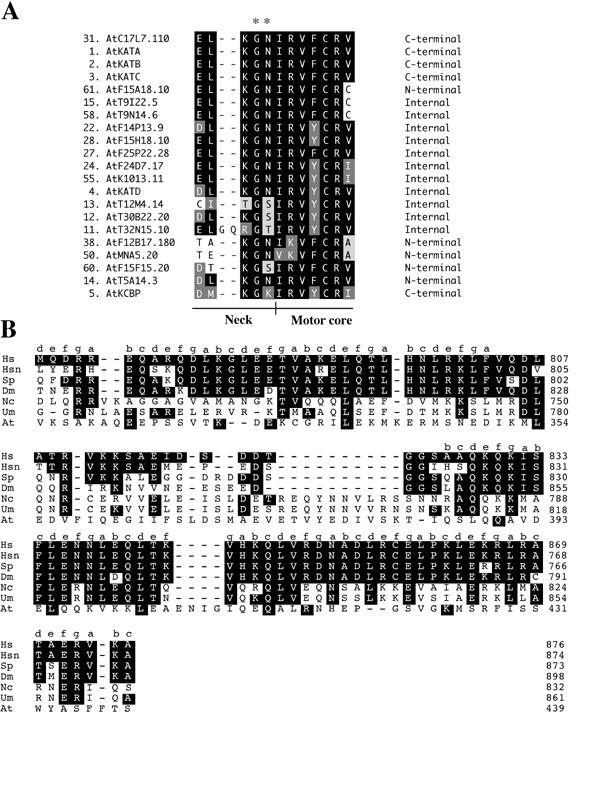
Figure 5.

Alignments of neck/motor core region and kinesin light chain binding site of KHC. Alignments were done using the Clustal method in DNA STAR MEGALIGN. A. Alignment of the neck/motor core regions from the 21 kinesins falling in the C-terminal subfamily. White letters on black are identical residues, white on dark gray are strongly similar and black on light gray are weakly similar. Asterisks mark the two residues shown to confer minus end directed movement [47]. B. Alignment of the kinesin light chain binding site in KHCs. The small letters indicate the heptad positions in the heptad repeats as given by Diefenbach et al. [52]. Hs, human KHC; Hsn, human neuronal KHC; Sp, sea urchin KHC, Dm, Drosophila melanogaster KHC; Nc, Neurospora crassa KHC; Um, Ustilago maydis, At, AtMAA21.110 kinesin.
