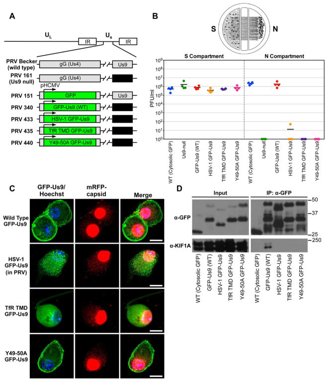
Figure 2. Characterization of PRV strains expressing wild type and functionally defective GFP-Us9 proteins.
(A) Schematic representation of the genomes of PRV Becker (wild type), PRV 161 (Us9 null), PRV 151 (diffusible GFP), PRV GFP-Us9 (wild type), PRV HSV-1 GFP-Us9 (HSV-1 Us9 in PRV), PRV TfR TMD GFP-Us9 (the transmembrane domain of Us9 was swapped for that of the transferrin receptor, thus making it non-lipid raft associated), PRV Y49-50A GFP-Us9 (mutated PRV Us9 in which the tyrosine residues at positions 49–50 have been substituted with alanines). The GFP and GFP-Us9 proteins are expressed ectopically from the HCMV promoter in the non-essential gG locus of the PRV genome. (B) Quantification of the efficiency of anterograde axonal spread using a chambered neuronal culture system. Cell bodies in the S compartment were infected at a high MOI with the indicated PRV strains. Four chambers were used for each viral strain. At 24 hpi, the entire content of the S and N compartments were harvested separately and titered on PK15 cells using a standard plaque assay. (C) Confocal microscopy of SCG neuron cell bodies infected with PRV strains expressing wild type and functionally defective GFP-Us9 proteins at 20 hpi (scale bars = 15 μm). (D) Western blot analysis of IPs from differentiated PC12 cells that are infected (at 20 hpi) with PRV strains that express the indicated GFP-tagged proteins. See also Figure S2 and Table S2.

