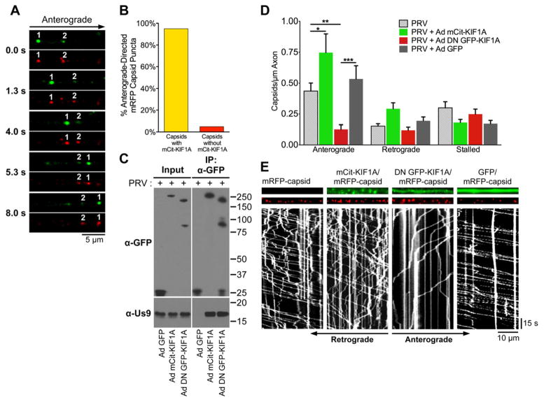
Figure 3. KIF1A mediates anterograde-directed transport of viral particles in axons.
(A) Live cell imaging of KIF1A (green) and capsid puncta (red) in axons of SCG neurons. Neurons were transduced with Ad mCit-KIF1A. At 4 days post transduction, neurons were infected with PRV mRFP-capsids and imaged between 8–12 hours post PRV infection. (B) Quantification of the percentage of anterograde-directed mRFP-tagged capsid puncta (mRFP-VP26) that co-transport with mCitrine-KIF1A in axons of SCG neurons. n = 555 capsids in 21 axons from 4 independent experiments. (C) PC12 cells were transduced with Ad GFP, Ad mCit-KIF1A, or Ad DN GFP-KIF1A and subsequently infected with PRV Becker. At 12 hpi (and 4 days post adenoviral transduction), cell lysates were prepared and subject to immunoaffinity purification using anti-GFP antibodies. (D) SCG neurons were transduced with Ad mCit-KIF1A, Ad DN GFP-KIF1A, Ad GFP, or mock. At 4 days post transduction, neurons were infected with PRV mRFP-capsids and visualized by live cell imaging between 8–12 hours post PRV infection. The number of anterograde, retrograde, and stalled particles was manually quantified and normalized to the measured axon segment length. Data is from: 12 axons containing 972 capsids for PRV mRFP-VP26 only; 9 axons containing 938 particles for Ad mCit-KIF1A and PRV mRFP-VP26, 14 axons containing 620 capsids for Ad DN GFP-KIF1A and PRV mRFP-VP26; 12 axons containing 999 capsids for Ad GFP and PRV mRFP-VP26. For each of these conditions, data was acquired from at least two independent replicates. Error bars indicate the mean ± SEM. * is p < 0.05, ** is p < 0.01, and *** is p < 0.001. (E) Representative kymographs of mRFP-capsids in axons of neurons transduced with the indicated adenoviral vectors. See also Movie S1.

