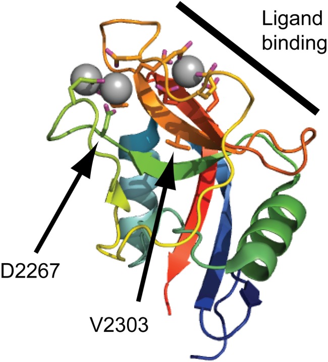
Figure 5.

Disease-linked missense mutations in the aggrecan C-type lectin domain (CLD). The aggrecan C-type lectin domain structure determined by X-ray crystallography (Protein Data Bank ID: 1TDQ) is shown as a cartoon model. The coordinated calcium ions are shown as gray spheres. The side chains of amino acid residues coordinating the calcium ions or mutated in human disease are shown as sticks. The CLD binding surface for tenascin-R is marked by a thick line in the upper right corner of the cartoon. The amino acid residues mutated in spondyloepimetaphyseal dysplasia aggrecan type (D2267) and familial osteochondritis dissecans (V2303) are indicated by arrows.
