Figure 1.

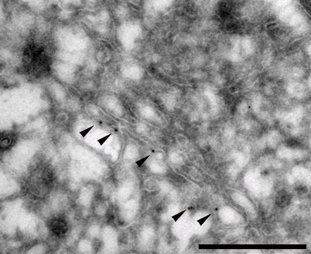
3′-Phosphoadenosine-5′-phosphosulfate transporter 1 (PAPST1) distribution in the Golgi apparatus of polarized cells. Madin-Darby canine kidney (MDCK II) cells expressing PAPST1–green fluorescent protein (GFP) were grown on polycarbonate filters for 4 days, fixed, and sectioned into 70-nm-thin sections in the cryomicrotome. Prior to examination by transmission electron microscope, the section was immunogold labeled using anti-GFP (Ab6556; Abcam, Cambridge, UK) and protein-A-Gold, followed by staining with uranyl acetate. Gold particles labeling PAPST1-GFP are observed at one side of the Golgi structure (black dots indicated by arrowheads). Scale bar = 500 nm.
