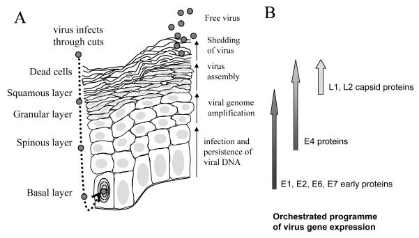
Figure 1. The replication cycle of high risk HPV in a differentiating epithelium.
A. The different epithelial layers are indicated on the left hand side of the diagram. Virus is show as small grey circles. The nucleus of the infected, dividing basal epithelial cell is indicated with curved lines representing condensed chromosomes. All nuclei are shaded in light grey. The granular layer is shown with dotted cytoplasm. The key events in the virus replication cycle are indicated to the right hand side of the diagram of the epithelium. B. A schematic diagram of the gene expression program of the virus within the infected epithelium. Shading on the arrows represents the quantity of expression of each protein subset during the virus replication cycle.

