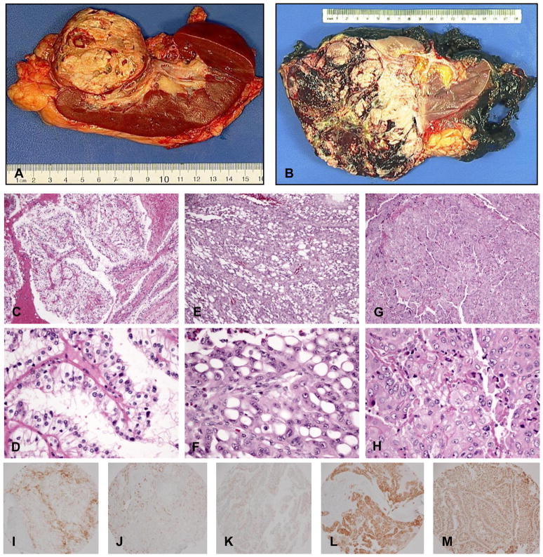
Figure 1.
Gross pictures, H&Es and IHC of Xp11 translocation RCC. (A), (B) Gross pictures from case #4 and #1, respectively. (C), (D) H&E from case #3 showed typical clear cells with papillary architecture. (E), (F) H&E from case #1 showed polygonal cells with large cytoplasmic vacuoles and eccentric nuclei (signet ring-like) with a microcystic growth pattern. (G), (H) H&E from case #5 showed a solid/syncytial growth pattern and grade 4 nuclear atypia. IHC (I) for CD10, (J) for AE1/3, (K) for Melan-A, (L) for HMB 45, and (M) for TFE3.

