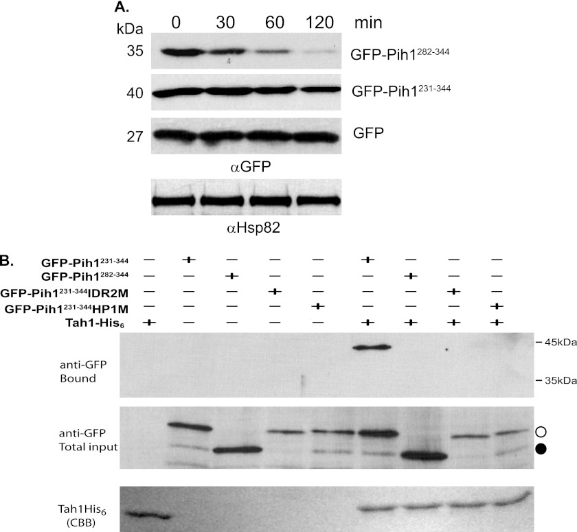
FIGURE 3.
Degradation of GFP Pih1 fusion proteins in vivo. A, degradation of GFP fusion proteins in yeast strain W303 after inhibition of protein translation using cycloheximide at 30 °C. Immunoblotting using anti-Hsp82 was used as an internal control. B, in vitro pulldown assays using His6-tagged Tah1 purified as bait from yeast cell lysates that express GFP-Pih1231–344, GFP-Pih1282–344, GFP-Pih1231–344IDR2M, or GFP-Pih1231–344HP1M. The top and middle panels are immunoblotting using anti-GFP antibody for the bound and total input fractions. The bottom panel shows Tah1 proteins bound to Ni-NTA. The open circle indicates the position of GFP-Pih1231–344 and its mutants. The closed circle indicates GFP-Pih1282–344.

