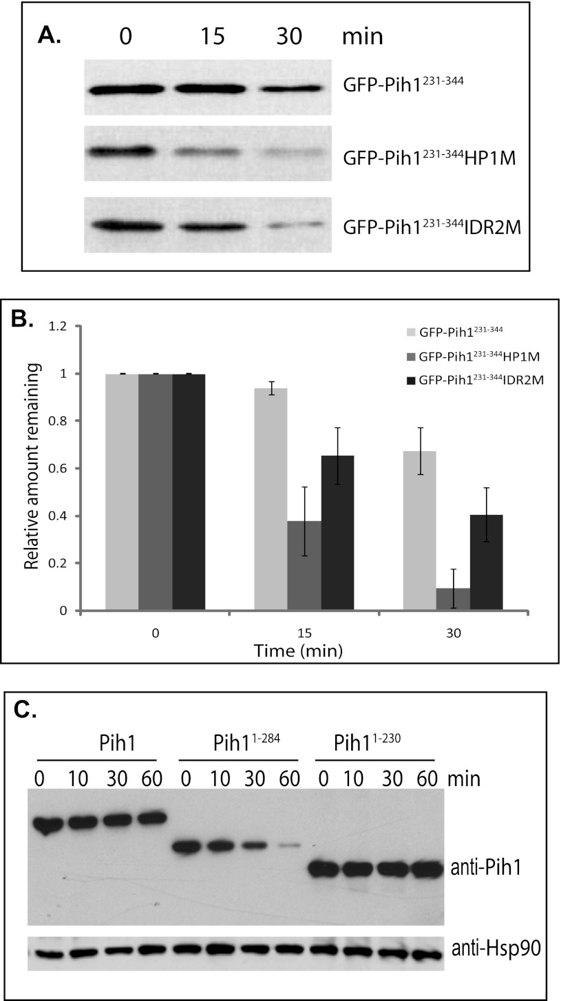
FIGURE 4.
Degradation of GFP Pih1231–344 fusion proteins and Pih1 truncation mutants in vivo. A, immunoblotting using anti-GFP antibody of GFP-Pih1231–344 fusion protein and its mutants expressed in yeast cells after inhibition of protein translation. B, quantitative analyses of GFP fusion protein degradation. The immunoblot was scanned and analyzed using Quantity One™ software (Bio-Rad) and the relative signal remaining was calculated by using the signal at 0 min as 100%. The averages and standard errors shown were from at least three independent degradation assays within 30 min following inhibition of protein translation with cycloheximide. C, immunoblotting of Pih1 and Pih1 truncation mutants Pih11–284 and Pih11–230 expressed in yeast cells which have their endogenous PIH1 genes deleted after inhibition of protein translation using cycloheximide. Immunoblotting using anti-Hsp82 was performed to detect Hsp90 expression and used as an internal control.

