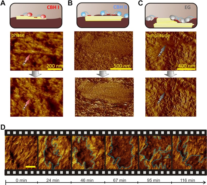
FIGURE 2.
Dissecting synergism: in situ observation of single enzymes. A, CBH I degrades small fibrils (phase image). B, CBH II polishes the surface of a large crystallite by removing amorphous cellulose (phase image). Right: (C), an example of EG polishing which leads to a highly defined fibrillar surface (amplitude image). D, “clustering” effect of CBH II in a phase image sequence (see also supplemental Video S3). The scale bar represents 100 nm.

