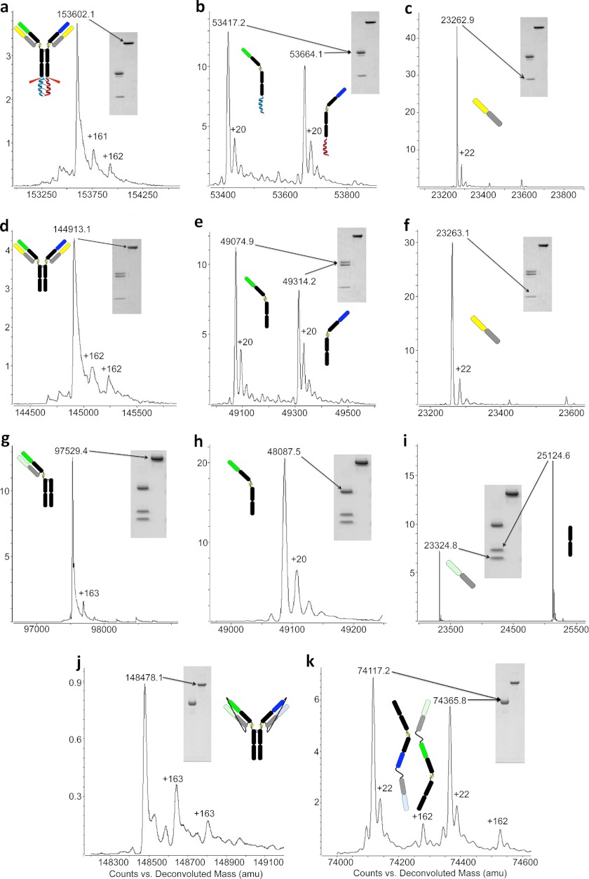
FIGURE 2.
Mass spectrometry and SDS-PAGE analysis of LUZ-Ys. a–c, common LC LUZ-Y with leucine zipper attached: non-reduced (a) and reduced yielding two HCs (b) and the common LC (c). d–f, common light chain LUZ-Y with leucine zipper removed non-reduced (d) and reduced (e and f). h and i, one-arm LUZ-Y with leucine zipper removed non-reduced (g) and reduced to yield HC (h), LC, and truncated Fc (i). j and k, tethered LUZ-Y with leucine zipper removed non-reduced (j) and reduced yielding two LC-HC-tethered antibody halves (k). A comparison of the experimentally determined masses to the theoretical masses is shown in supplemental Table 1.

