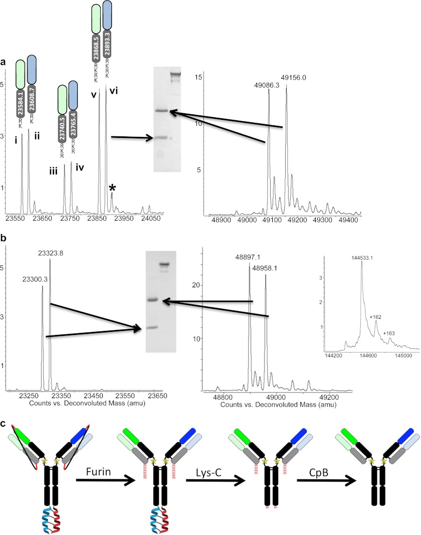
FIGURE 3.
Anti-HER2/EGFR LUZ-Y with furin cleavable tethers. a, SDS-PAGE and MS analysis of anti-HER2/EGFR LUZ-Y LCs (containing residual furin site residues at the C terminus) and HCs following purification and Lys-C processing to remove the leucine zippers. Three major species of each LC are present after initial purification steps of the LUZ-Y Ab; LC + RK (i and ii), LC + RKR (iii and iv), and LC + RKRK (v and vi). b, SDS-PAGE and MS analysis of the anti-HER2/EGFR LUZ-Y following carboxypeptidase B treatment to remove the residual furin site from the C terminus of the light chains. Inset shows mass spectra for the non-reduced intact antibody post carboxypeptidase B (CpB) treatment. c, schematic representation of the overall process. *, additional mass is the LC+RKRK plus a sodium ion.

