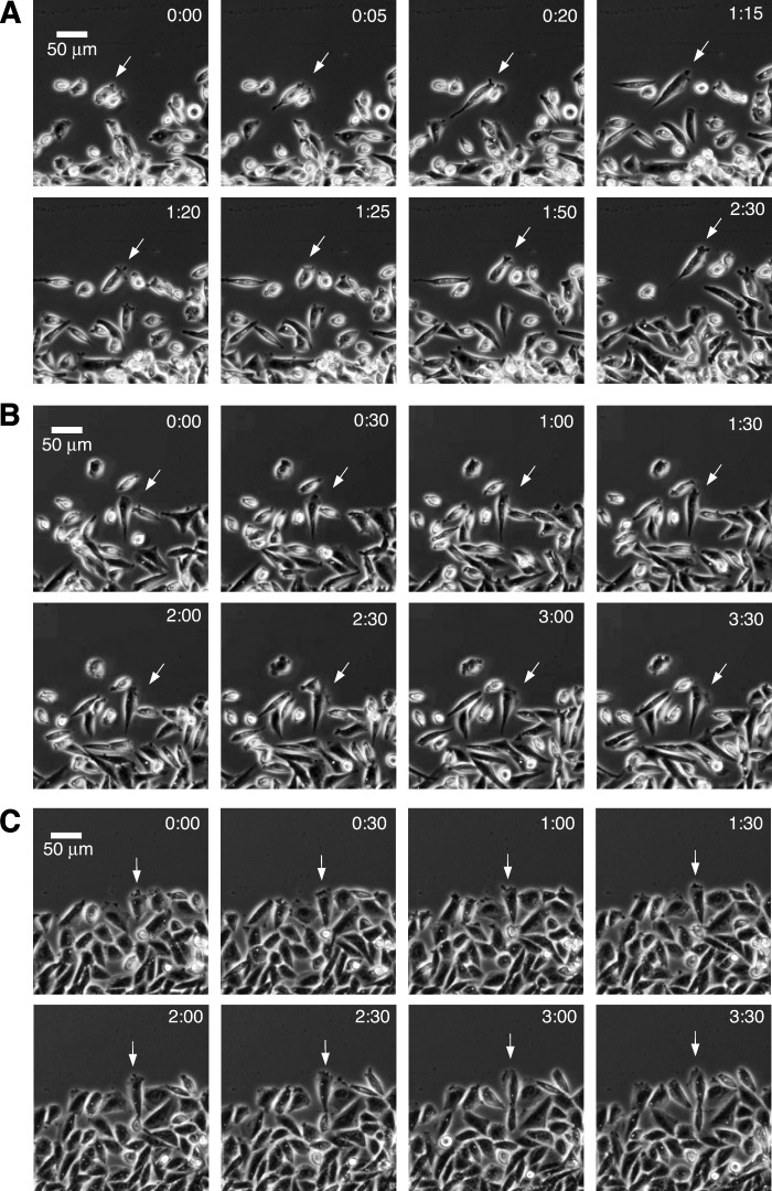
FIGURE 4.
Time-lapse video of cells moving into a wound. Wild-type (A), wild-type treated with 5 nm paclitaxel (B), and mutant CV 2-8 (C) are shown. The cells were photographed with a 10× objective every 1 min using phase contrast optics. The time in h:min from the start of the sequence is indicated. Scale bar, 50 μm. The arrows were added to aid in tracking individual cells through the time lapse sequences.

