
FIGURE 4.
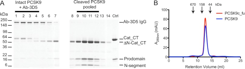
Purification and analysis of furin-cleaved PCSK9. A, PCSK9 was treated with furin for 20 h, after which an anti-furin antibody and Ab-3D5 were added, and the protein mixture was applied to a quantitative S-200 size exclusion column. The first elution peak contained the complex formed of Ab-3D5 with residual intact PCSK9 (∼60-kDa Cat_CT; lanes 1–4) and was separated from the later eluting pure cleaved PCSK9 (∼50-kDa ΔN-Cat_CT; lanes 9–12) (see also Suppl. Fig. 4). B, pooled fractions (9–12) of pure furin-cleaved PCSK9 (PCSK9c_fu; red elution profile) were compared with intact PCSK9 (PCSK9; blue elution profile) by analytical size exclusion chromatography. The elution volumes for PCSK9c_fu and PCSK9 were identical (12.72 and 12.78 ml), both having a deduced mass of 77 kDa. Molecular mass markers are indicated by arrows.