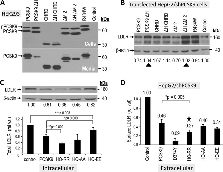
FIGURE 6.
Impact of basic residues in the hinge region and M2 module on the activity of PCSK9. Hinge region: A, HepG2/shPCSK9 cells were transiently transfected with various V5-tagged constructs containing or lacking the hinge region of PCSK9 (aa 405–420) and/or lacking the M2 module. At 48 h post-transfection, the proteins in the media and cell lysates were separated by SDS-PAGE and analyzed by Western blot using mAb-V5. The migration positions of proPCSK9 (pPCSK9) and mature PCSK9 are emphasized. B, the total LDLR-degrading activity of each construct was analyzed 48 h post transfection by Western blot of cell lysates using a polyclonal LDLR antibody, and the levels of the ∼160 kDa were normalized to β-actin cellular loading controls. The ∼110 kDa LDLR represents the immature form of the receptor. The black triangles point to the abrogation of the intracellular activity in WT and ΔM2 constructs upon removal of the hinge region. M2 module: C, HepG2/shPCSK9 cells were transiently transfected with various V5-tagged constructs expressing PCSK9 ΔM2, WT PCSK9 or some of its single point mutant (GOF D374Y) and the charge modified double point mutants (HQ553,554RR; HQ553,554AA; HQ553,554EE). The total LDLR-degrading activity of each construct was analyzed in B. D, cell surface LDLR levels were evaluated by FACS analysis following 4 h incubation of HepG2/shPCSK9 cells with most of the constructs in C. The star emphasizes the GOF of the HQ553,554RR mutant in this assay. These data are representative of at least three independent experiments. Statistical significance is emphasized (*, p < 0.05).

