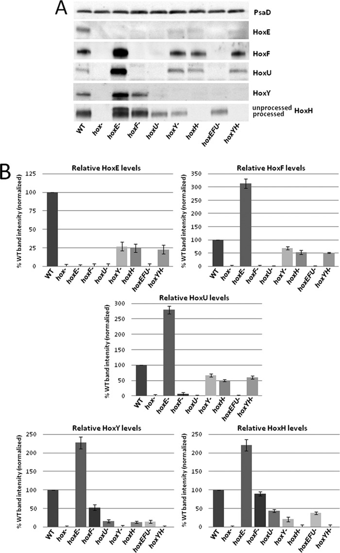
FIGURE 3.
One-dimensional SDS-PAGE and Western blot analysis of Synechocystis sp. PCC 6803 hox mutants. A, whole cell lysates of WT and individual/combined hox mutants were run on TGX Any kDa gradient SDS-polyacrylamide gels (Bio-Rad), transferred to PVDF, and immunoblotted with Hox subunit-specific antibodies and PsaD and/or Rps1 (not shown) as loading controls. B, relative levels of Hox subunits in each mutant, presented as a percentage of WT. Data represent quantitation of multiple Western blot analyses with error bars depicting variation between separate analyses.

