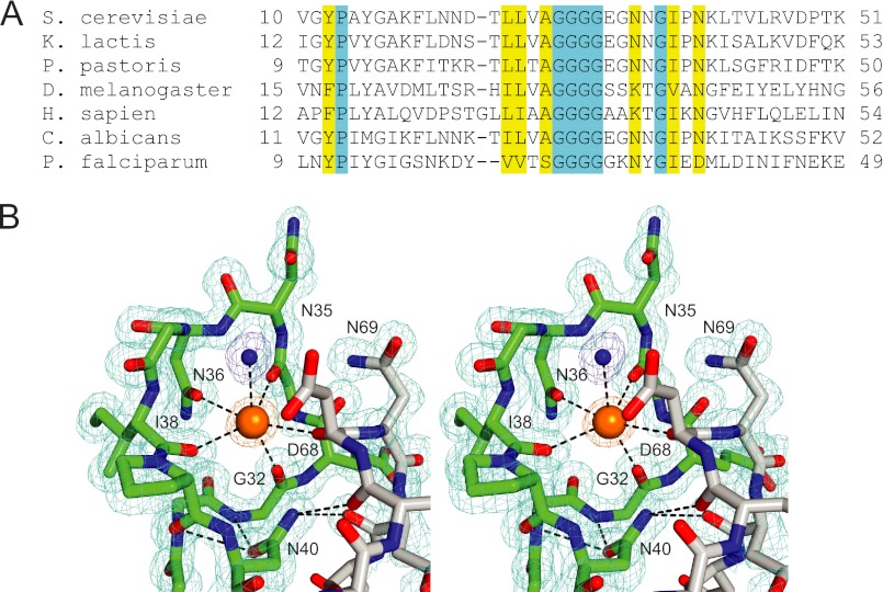
FIGURE 2.
The K loop region of Sec12. A, sequence alignment. Much less homology is observed outside the K loop region of the cytoplasmic domain, and virtually no homology is observed in the luminal domain. B, stereo view of the K loop (green), adjacent loop (gray), and corresponding electron density (2Fo − Fc, contoured to 1σ). The bound K+ and liganding water molecule are shown as a large orange sphere and a small blue sphere, respectively. The residues that contribute the remaining five oxygen ligands (except for Gly-34) are labeled. Also displayed is the hydrogen-bonding network centered on the functionally critical side chain of Asn-40.

