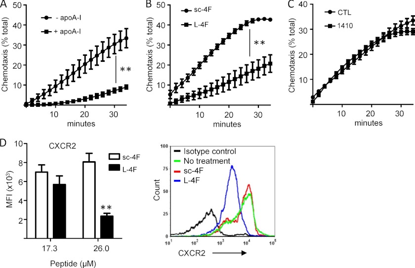
FIGURE 4.
Apolipoprotein mimetics suppress CXCR2-directed PMN chemotaxis. A-C, wild type murine bone marrow-derived PMNs were pretreated for 1 h with apoA-I (20 μg/ml) or media control (A), L-4F or sc-4F (10 μg/ml) (B), or COG1410 or control (CTL) peptide 264 (8.5 μg/ml) (C), and then assayed for chemotaxis (represented as % of total input PMNs) up a MIP-2 gradient. Data are mean ± S.E. and represent 2 or more independent experiments (**, p < 0.01). D, cell surface display of CXCR2 by murine PMNs was quantified by flow cytometric measurement of CXCR2 mean fluorescence intensity (MFI) after cell treatment (1 h) ex vivo with the indicated concentrations of sc-4F or L-4F. Data are mean ± S.E. and represent 2 independent experiments; **, p = 0.001. A representative flow cytometry histogram is shown at right.

