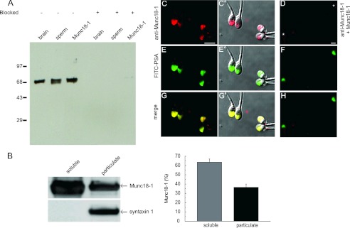
FIGURE 1.
Munc18-1 is present in human sperm. A, postnuclear membrane pellet from rat brain (1 μg of protein, brain), a human sperm extract (5 × 10⁶ cells corresponding to 5 μg of protein, sperm), and recombinant Munc18-1 (Munc18-1, 1 ng) were resolved in 10% Tricine gels, transferred to nitrocellulose membranes, and probed with an anti-Munc18-1 antibody from Synaptic Systems (left) or the antibody preblocked with the recombinant protein (4-fold molar excess, right). Molecular weight markers ( × 10−3) are indicated on the left. B, sperm disrupted by nitrogen cavitation and separated into particulate and soluble fractions by ultracentrifugation were loaded (5 × 10⁶ cells) onto SDS-polyacrylamide gels and probed on blots with the anti-Munc18-1 (top) and anti-syntaxin 1 (bottom) antibodies from Abcam (left). The percentage in each fraction was quantified in three independent experiments; the mean ± S.E. is shown on the right. C–H, sperm were fixed on coverslips and labeled with the anti-Munc18-1 antibody from Synaptic Systems. The acrosomal region was stained with fluorescein isothiocyanate-coupled PSA-FITC that recognizes the intra-acrosomal content. Shown are epifluorescence micrographs of typically stained cells with the anti-Munc18-1 antibody (C) and anti-Munc18-1 antibody preblocked with 4-fold excess recombinant protein (D), PSA-FITC (E and F), and colocalization (G and H). In C′, E′, and G′, the fluorescence images were superimposed to differential interference contrast (DIC) images to depict the localization of the labels within the cells. Notice that Munc18-1 localized to the acrosomal region. Bar, 5 μm. Asterisks in D indicate cells with intact acrosomes but without Munc18-1 staining.

