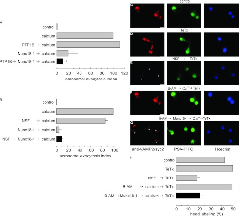
FIGURE 5.
Recombinant Munc18-1 halts acrosomal exocytosis at a stage when SNAREs are monomeric. A and B, SLO-permeabilized spermatozoa were loaded with 27 nm PTP1B (A) or 300 nm NSF (B) and incubated for 15 min at 37 °C to disassemble cis-SNARE complexes. Next, we added 4 nm Munc18-1 and incubated for a further 15 min at 37 °C. Finally, we added 0.5 mm CaCl2 and incubated for an additional 15 min to stimulate exocytosis (black bars). Controls (gray bars) included the following: background exocytosis in the absence of any stimulation (control); exocytosis stimulated by 0.5 mm CaCl2 (calcium); exocytosis inhibited by 4 nm Munc18-1 and unperturbed by 27 nm PTP1B or 300 nm NSF. Sperm were fixed, and acrosomal exocytosis was evaluated using PSA-FITC, and data normalized as described under “Experimental Procedures.” The values represent mean ± S.E. of at least three independent experiments. C–G, we incubated SLO-permeabilized sperm under resting conditions (C and D) or with 300 nm NSF to promote cis-SNARE complex disassembly (E). At the end of the incubation, we added 100 nm light chain of TeTx to test VAMP2/syb2 neurotoxin sensitivity (D and E). Other aliquots were incubated with 10 μm BAPTA-AM (B-AM, F and G). When indicated, 4 nm Munc18-1 was added to the assay (G). Next, we added 0.5 mm CaCl2 to stimulate exocytosis. At the end of the incubation, we added 100 nm light chain of TeTx. We then fixed and triple-stained the cells with an anti-VAMP2/syb2 antibody (red, left panels), PSA-FITC (to differentiate between reacted and intact sperm; green, central panels), and Hoechst 33342 (to visualize all cells in the field; blue, right panels). Asterisks indicate cells with intact acrosomes but without VAMP2/syb2 immunostaining due to toxin cleavage. Bars, 5 μm. H, quantification of the percentage of cells treated as in C–G exhibiting VAMP2/syb2 acrosomal staining from three independent experiments (mean ± S.E.).

