
FIGURE 5.
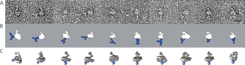
Electron microscopy of the purified DHPR complex bound to anti II-III loop antibody. A, raw images of the L-type channel labeled with anti-α1s-II-III loop antibody. B, schematic representation of the antibody (blue) labeling the DHPR complex (white) in the orientation found in A. C, 3D reconstruction of the DHPR in the corresponding orientation. The blue sphere represents the location found for the antibody. Scale bar = 10 nm.