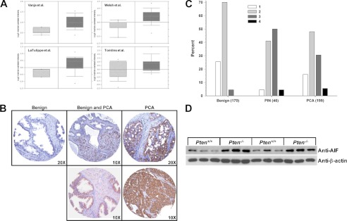
FIGURE 1.
AIF is elevated in prostate cancer. A, AIF mRNA levels as assessed in four representative studies archived in the Oncomine cancer gene expression analysis database (28–30, 32). Light gray bars, AIF expression in normal prostate tissue; dark gray bars, AIF expression in prostate cancer. B, immunohistochemistry was performed on tissue microarrays derived from samples of benign (left panel), a mixture of benign and prostate carcinoma (center panels), and frank carcinoma (right panels). AIF staining is shown in brown, and hematoxylin staining is shown in blue. C, microarray samples classified as benign, prostatic intraepithelial neoplasia (PIN), or frank carcinoma (PCA) were scored for AIF staining intensity on a scale of 1–4. Note the higher proportion of samples with intense AIF staining (score 3 or 4) present in PIN and PCA samples (p < 0.0001 by Kruskal-Wallis test). D, prostate tissue was harvested from six wild type (Pten+/+) and six prostate-specific Pten-deficient (Pten−/−) mice. Protein was extracted, and AIF levels were determined by immunoblot analysis. As a loading control, extracts were immunoblotted for β-actin levels.

