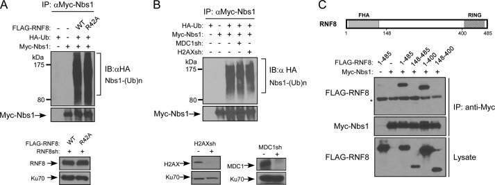
FIGURE 3.
RNF8-mediated ubiquitination of Nbs1 is independent of MDC1. A, in vivo ubiquitination assay. U2OS cells stably expressing FLAG-tagged RNF8-WT or RNF8-R42A FHA mutant, with endogenous RNF8 silenced by shRNAs, were transfected with Myc-Nbs1 and HA-ubiquitin. 40 h later, cells were lysed, immunoprecipitated with anti-Myc antibody, and then probed with anti-HA antibody for revealing ubiquitinated Nbs1. B, in vivo ubiquitination assay. U2OS cells were transfected with Myc-Nbs1 and HA-ubiquitin and then retrovirally infected with or without MDC1 or H2AX shRNA viruses. 40 h later, cells were lysed, immunoprecipitated with anti-Myc antibody, and then probed with anti-HA antibody for showing ubiquitinated Nbs1. Immunoblots show silencing of H2AX and MDC1, with Ku70 used as a loading control. C, top, schematic drawing of RNF8 protein domain structure, including FHA and RING domains. Bottom, in vivo ubiquitination assay of 293T cells transfected with Myc-Nbs1 or FLAG-tagged RNF8 (full-length 1–485, N-terminal deletion 148–485, C-terminal deletion 1–400, or combined N-terminal and C-terminal deletion 148–400) plasmids. Cells were lysed and immunoprecipitated with anti-Myc antibody, followed by immunoblotting with the indicated antibodies. *, nonspecific band. IB, immunoblot.

