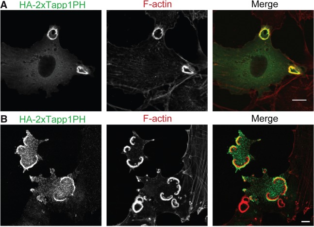
Fig. 3.
Localizations of PI(3,4)P2 at CDRs and podosome rosettes. (A) NIH3T3 cells expressing HA-2 × Tapp1PH [a specific probe for PI(3,4)P2] were stimulated with PDGF for 5 min, and then stained with anti-HA antibodies as well as rhodamine-phalloidin. (B) NIH3T3 cells expressing an active form of Src (Y530F) were transfected with HA-2 × Tapp1PH, and then stained with anti-HA antibodies as well as rhodamine-phalloidin. Bars, 10 µm.

