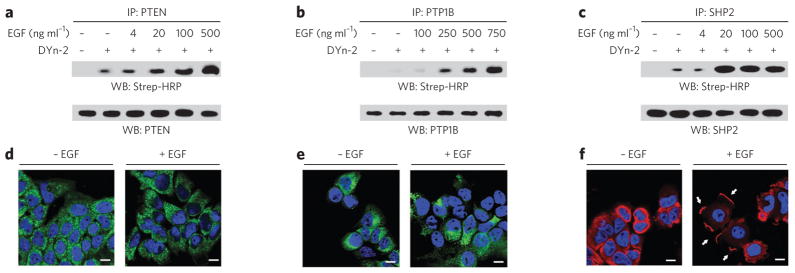
Figure 4. Differential sulfenylation of PTPs in EGF-treated cells.
(a–c) Western blots (WB) showing sulfenylated and total immunoprecipitated PTEN, PTP1B and SHP2. A431 cells were stimulated with EGF or vehicle for 2 min at the indicated concentrations and then incubated with 5 mM DYn-2 or vehicle for 1 h at 37 °C. Lysates were immunoprecipitated with mouse PTEN- (a), mouse PTP1B- (b) or rabbit SHP2-specific antibody (c). Sulfenylation of PTPs was detected by Strep-HRP western blot. Western blots were reprobed for total PTP as indicated to verify equivalent recovery. (d–f) Confocal fluorescence images of A431 cells stimulated with vehicle or 100 ng ml−1 EGF for 5 min. Cells were stained with PTEN- (d), PTP1B- (e) or SHP2-specific antibody (f). Nuclei were counterstained with DAPI (blue). Scale bars, 10 μm. The white arrows in f highlight the changes in subcellular localization of SHP2 after stimulation with EGF.

