
Figure 2. Carcinoma-derived CXCL1/2 supports cancer cell survival and recruit granulocytic myeloid cells to tumors.
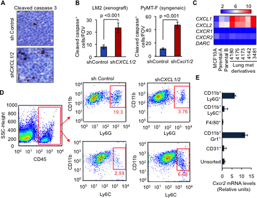
(A–B) Representative images and quantification of apoptosis in mammary tumors analyzed by Cleaved caspase-3 staining. Mouse mammary glands were injected with LM2 cells (A,B) or PyMT-F cells (B) expressing shRNA control or shCXCL1/2 and analyzed at endpoint (LM2, 6 weeks; PyMT-F, 9 weeks after tumor implantation). Scale bar equals 30µm. Data are averages ± SEM. n=4 mice per group. P values were calculated by Student’s t-test.
(C) Expression of the indicated genes from microarray gene expression analysis in non-tumor human mammary epithelial cell line MCF10A, parental MDA-MB-231 breast cancer cells and lung metastatic lines derived from MDA-MB-231 (Minn et al., 2005).
(D) Flow cytometric analysis of recruited myeloid cells in tumors formed by LM2 cells transduced with either control shRNA or shCXCL1/2 at 5 weeks after tumor inoculation. A representative gating is shown. Numbers indicate either CD11b+Ly6G+ or CD11b+Ly6C+ cells in the quadrant expressed as percentages of total CD45+ leukocytes from the same tumor. Results are representative of three independent experiments (n=3).
(E) Expression of Cxcr2 receptor in sorted subpopulations of LM2 tumors determined by qRT-PCR. Error bars represent 95% confidence interval for qRT-PCR analysis. Data is representative of two independent experiments.
See also Figure S2 and Tables S1 and S2