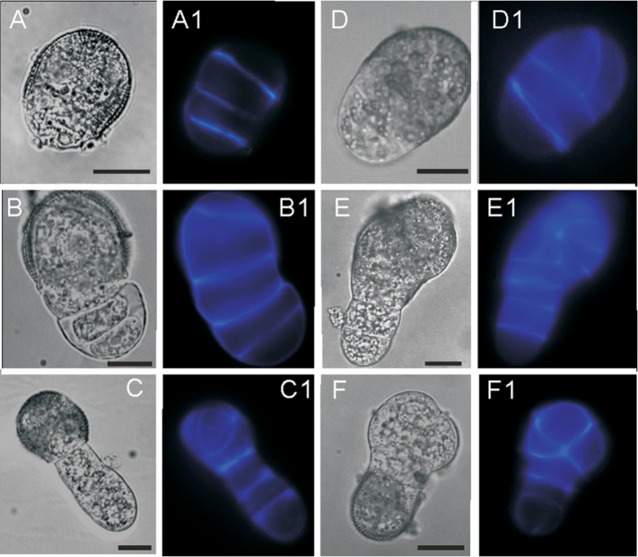
Fig. 3.
Division pattern of the EDM-derived suspensors. (A–F) Bright field images. (A1–F1) Fluorescence microscopic images. Cells were stained with CW. (A–C1) Both daughter cells of the EDM underwent transverse divisions at first. The exine-covered cell underwent several divisions and developed into an embryo proper, while the naked cell kept transverse division and developed into a suspensor. (D–E1) The exine-covered cell underwent longitudinal cell division and later developed into an embryo proper. The naked cell kept transverse division and developed into a suspensor. (F, F1) The exine-covered cell divided transversely, giving rise to a suspensor. Bars=5 µm.

