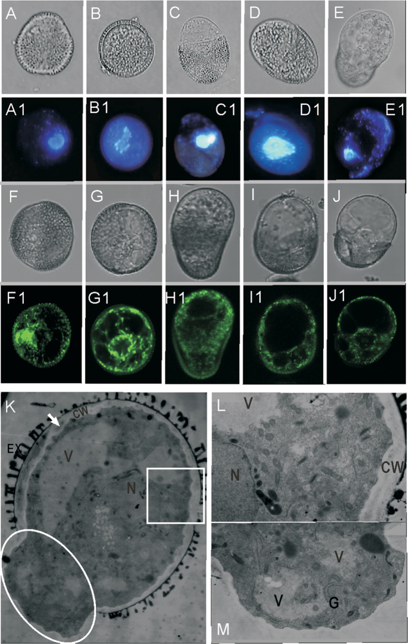
Fig. 4.
Cytological characters of EDMs. (A–J) Light microscope images. (A1–J1) Fluorescence microscope images. (A, A1) An exine-intact microspore, showing that the nucleus locates near the pollen wall. (B, B1) An exine-intact microspore after heat shock, showing that the nucleus is located centrically. (C–E1) The nucleus locates at the plane between the exine-covered part and the exine-detached part. Organelle DNA is usually distributed around the nucleus in all type 1 (D, D1), type 2 (E, E1), and type 3 EDMs. (F, F1) An exine-intact microspore, showing that mitochondria distribute near the pollen wall. (G, G1) A exine-intact microspore after heat shock, showing that mitochondria are located centrically around the nucleus. (H–J1) Polar distribution of mitochondria in all type 1 (I, I1), type 2 (J, J1), and types 3 EDMs. Bars=5 µm. (K–M) Ultrastructural observation of EDMs. (K) Low magnification micrograph showing the entire cytological features; the intine became much thicker on the exine-covered side of the EDM. (L) Amplifyication of the boxed part in K, showing more organelles, especially mitochondria, in the exine-covered part. (M) Amplification of the ellipsoid part in K, showing more vacuolation and more endoplasmic reticulum in the naked part. EX, exine; CW, cell wall; V, vacuole; G, Golgi body; N, nucleus. Bars=0.5 µm.

