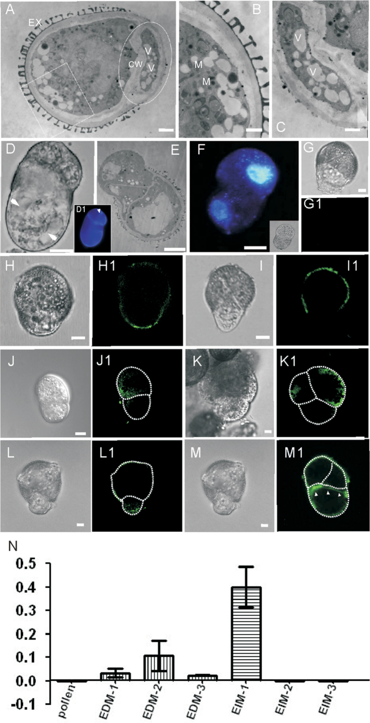
Fig. 6.
Comparisons between two daughter cells of EDMs. (A–C) Ultrastructural observations of a divided EDM. (A) Low magnification micrograph shows the entire cytological features. (B) Amplification of the square area in (A); the larger cell has condensed cytoplasm and more mitochondria. (C) Amplification of the ellipsoid area in (A), showing that the small cell is more vacuolated. EX, exine; CW, cell wall; V, vacuole; M, mitochondria. Bars=0.5 µm. (D) Vacuoles were observed in the larger naked cell after asymmetric cell division; the arrows indicate vacuoles. (D1) CW staining of the cell in (D). The arrowhead indicates the division plane. (E) Thin section of a symmetrically divided EDM, showing that an exine-covered cell contains a large vacuole. (F) A symmetrically divided EDM (indertion), showing that organelle DNA differentially distributes in its two daughter cells. (G–I1) Immunolocalization of JIM8. (G, G1) Control, showing no fluorescence on the exine. (H, H1) JIM8 locates more on the small daughter cell. (I, I1) JIM8 locates more on the large daughter cell. (J, J1) PIN1 mainly localizes in the large daughter cell. (K, K1) The cell with PIN1 signal divided longitudinally and will probably develop into an embryo proper. (L, L1) PIN7 appears in the small daughter cell, which will probably develop into a suspensor. (M, M1) The cell without PIN7 signal divided vertically. (N) BnWOX8 expression level in different microspore embryos. (EDM-1) The first divided EDMs. (EDM-2) Small embryo with suspensor (4 µm in diameter). (EDM-3) Large embryo with suspensor (12 µm in diameter). EIM, exine-intact microspores. EIM1, the first divided EIM; EIM2, small globular embryos without a suspensor (4 µm in diameter); EIM3, large microspore embryos without a suspensor (12 µm in diameter). Data are the mean of three independent experiments. Bars indicate standard errors.

