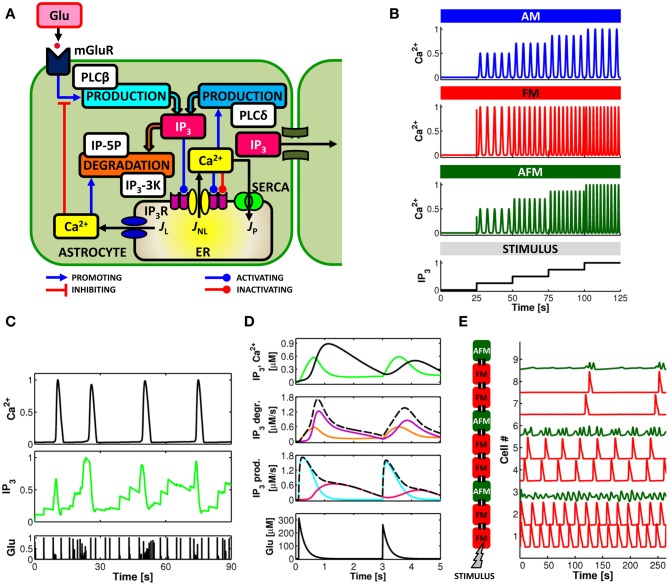
Figure 2.
Computational aspects of astrocytic Ca2+ signaling. (A) Scheme of IP3-mediated Ca2+-induced Ca2+ release in the astrocyte. Calcium and IP3 signals are controlled by synaptic glutamate through metabotropic glutamate receptor- (mGluR-) PLCβ-mediated IP3 production (see text for details). (B) IP3 and Ca2+ signals can be envisioned to encode incoming synaptic activity through frequency and amplitude of their oscillations. Astrocytes could thus encode synaptic information either by modulations of the amplitude (AM), the frequency (FM), or both (AFM) of their Ca2+ oscillations. (C) Simulated Ca2+ and IP3 patterns in response to sample synaptic glutamate release (Glu) in a model astrocyte (De Pittà et al., 2009a,b) reveals that IP3 signals could be locked in the AFM-encoding independently of the encoding mode of the associated Ca2+ signals. This feature could allow the astrocyte to optimally integrate synaptic stimuli. (D, top) Simulated Ca2+ (black trace) and IP3 (green trace) signals in the same astrocyte model as in (C), and associated rates of IP3 production (prod.) and degradation (degr.) (middle panels, dashed black lines) in response to two consecutive synaptic glutamate release events (bottom panel, Glu). The analysis of the contributions of different enzymes to IP3 signaling (solid colored traces; cyan: PLCβ ; pink: PLCδ ; orange: IP-5P; and purple: IP3-3K) reveals dynamical regulation by Ca2+ of different mechanisms of IP3 production/degradation which could ultimately underlie dynamical regulation of astrocyte processing of synaptic stimuli. (E) Simulated propagation of Ca2+ waves in a heterogeneous linear chain composed of both FM (red traces) and AFM (green traces) astrocytes reveals that encoding of synaptic activity (STIMULUS) could change according to cell location along the chain. Adapted from Goldberg et al. (2010).

