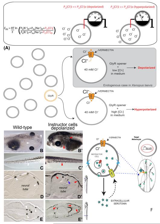
Figure 1.
Depolarization of instructor cells induces metastatic phenotype in melanocytes.
The Goldman equation reveals how resting potential Vmem depends on the internal and external K+, Na+ and Cl− concentrations, ambient temperature (T), and permeability of each ion (P).
(A) Manipulation of endogenous chloride channels as a means for selective probing of Vmem-based signaling. Ivermectin treatment targets GlyCl-expressing (“instructor”) cells, opening these chloride channels. Then, by manipulating external chloride levels, chloride ions can be made to enter or exit the GlyCl-expressing cells, thus controlling their transmembrane potential.
Normal tadpoles exhibit small numbers of black melanocytes over their neural tubes but not around their eyes or mouth (B, white arrowheads). In contrast, tadpoles whose instructor cells were depolarized for as little as 12 hours exhibit excess melanocytes, which possess a much more arborized appearance, and colonize areas normally devoid of melanocytes (B’, red arrowheads). The same change in shape is seen in the tail (compare normal tail in panel C with the tail of a depolarized tadpole in C’); this change was quantified and mitotic indices were calculated in (Blackiston et al., 2011b).
Sectioning of these tadpoles reveals that in contrast to the small number of round melanocytes normally present in the neural tube (D, white arrowheads), depolarization of instructor cells causes melanocytes to acquire a highly aberrant spread-out morphology (D, red arrowheads) as well as to invade the nervous tissue (blue arrowhead). This is also seen in more posterior sections through the tail/trunk, where the normally round melanocytes (E, white arrowheads) are far more numerous and extend long abnormal projections (E’, red arrowheads).
(F) Because the phenotype can be recapitulated by gain-of-function of serotonin signaling, and prevented by blockade of the serotonin transporter SERT, the data suggest a model of how instructor-cell signaling affects melanocyte morphology: depolarized instructor cells’ SERT runs backwards, secreting serotonin into the extracellular space where it can diffuse and activate melanocytes to acquire 3 basic properties of metastasis: overgrowth, abnormal cell shape, and invasion of other tissues.

