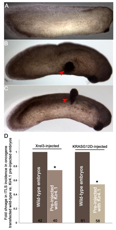
Figure 7.
Oncogene-induced ITLSs can be suppressed by prior injection of hyperpolarizing channel mRNA.
(A) An unperturbed (control) stage 29 embryo showing normal development.
(B) Embryos that are electroporated with Xrel3 DNA at stage 10 exhibit ITLSs by stage 29 (red arrowhead).
(C) Embryos that are electroporated with KRASG12D DNA at stage 10 exhibit ITLSs by stage 30 (red arrowhead).
(D) Xrel3 and KRASG12D ITLS formation can be partially blocked by forced pre-hyperpolarization via molecular expression of Kir4.1 (a hyperpolarizing channel). Injection of Kir4.1 mRNA at the one-cell stage followed by electroporating with oncogene DNA at stage 10 results in 25.8% fewer embryos with ITLSs compared with Xrel3 electroporation on a wild-type background and 46% fewer embryos with ITLSs compared with KRASG12D electroporation on a wild-type background. *p<0.05 One-sample t-test to a normalized ITLS incidence (set to 1) in Xrel3- or KRASG12D-electroporated embryos.

