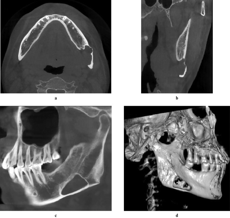
Figure 2.
Axial (a), coronal (b) and sagittal (c) sections of cone beam CT (CBCT) showing a large bone cavity with an irregular border. The inner tissue seemed to be continuous with the submandibular gland. The buccal cortex was expanded and perforated in three-dimensional volume rendering processing (d)

