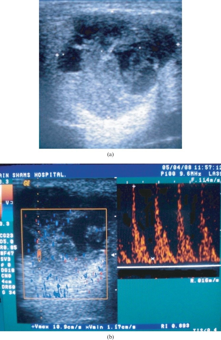
Figure 4.
(a) Greyscale sonography (GSS) of an enlarged right parotid gland showing a well-defined, hypoechoic, heterogeneous lesion with multiple anechoic areas and a solid component. (b) Colour Doppler sonography (CDS) of the same case showing Grade 3 scattered vascularity, spectral Doppler (SPD) showing peak systolic velocity of 10.9 cm s−1 (below the threshold level), high resistive index of 0.89 (above the threshold level), Vmin of 1.17 cm s−1 so pulsatility index = 1.6 (above the threshold level). Histopathological examination revealed the lesion to be mucoepidermoid carcinoma

