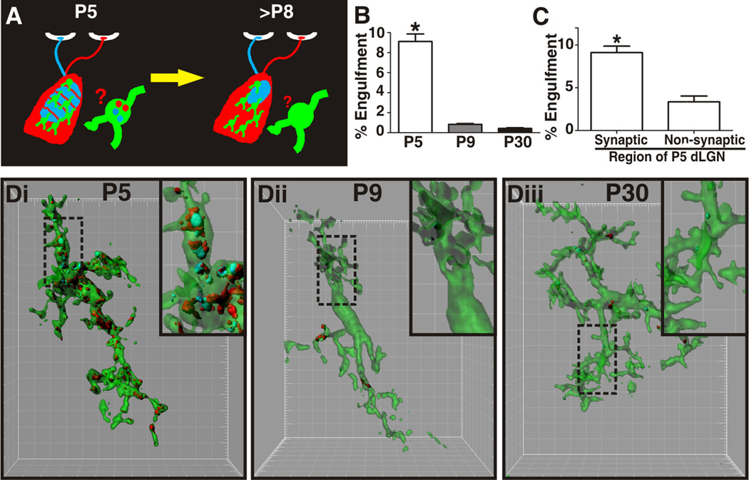
Figure 2. Microglia-mediated engulfment of RGC inputs is developmentally regulated.
A, Schematic of retinogeniculate pruning and strategy used for assessing engulfment. Contralateral (red) and ipsilateral (blue) inputs overlap at early postnatal ages (P5). Inputs from both eyes prune throughout the dLGN during the first postnatal week and is largely complete by P9/10. Engulfment was analyzed throughout the dLGN. B, Engulfment of RGC inputs is significantly increased during peak pruning in the dLGN (P5). *P<0.001 by one-way ANOVA, n=3 mice/age. C, Engulfment in P5 dLGN occurs most significantly in synapse-enriched (contralateral and ipsilateral dLGN) versus non-synaptic (optic tract) regions. *P<0.01 by Student’s t-test, n=3 P5 mice. All error bars represent s.e.m. D, Representative surface rendered microglia from P5 (fluorescent image is shown in 1B), P9, and P30 mouse dLGN. Engulfment of RGC inputs occurs during peak pruning (P5) versus older ages (P9 and P30). Enlarged insets denoted with a black dotted line. Grid line increments = 5 µm. See also Figure S2.

