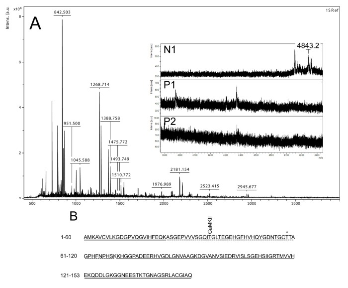
Figure 3.
Mass spectrometry analysis of spot N1, P1 and P2. (A) MALDI-MS spectrum of tryptic peptides obtained from P1 spot shown in Figure 2. N1, P1, and P2 were all identified as SOD1. Mass spectra were similar for all three spots (N1, P1 and P2), except that the long tryptic peptide (4843.2 m/z), corresponding to aa 24–69, was only observed in N1 (insert of A). Sequence covered by all three spots is underlined in (B). Dotted line refers to sequence covered only in N1 and represents the long tryptic peptide aa 24–69. This peptide has several possible Ser/Thr phosphorylation sites including a consensus sequence site for CaMKII and also a previously suggested phosphorylation site (asterisk) (Thr58) [16].

