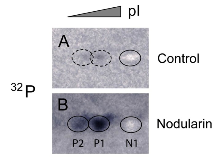
Figure 4.

Immunoprecipitation of SOD1 from 32P-labeled apoptotic hepatocytes. Hepatocytes were pre-labeled with 32Pi and exposed to 5 µM nodularin for 2 min. Total cell extract was immunoprecipitated with anti-SOD1 antibody and separated by two-dimensional gel electrophoresis (pI 4–7, 7 cm). See Experimental Section for further details. The figure shows autoradiographs from control (A) and nodularin-treated (B) cells. The non-phosphorylated silver-stained SOD1 (spot N1) appeared as a white spot on the autoradiographs in both control- and nodularin-treated hepatocytes. Nodularin treatment led to increased abundance of the two phosphorylated SOD1 spots (P1 and P2). The effect was more pronounced than observed with a lower concentration of nodularin (Figure 2).
