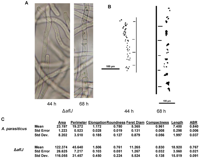
Figure 6.
Comparison of vesicle/endosome distribution in the A. parasiticus ΔaflJ mutant grown in the dark for 44 h and 68 h on YES liquid medium. (A) Morphology of ΔaflJ was evaluated at indicated time points using bright field microscopy; (B) Binary images of the corresponding distribution of vesicles and endosomes in wild-type A. parasiticus and ΔaflJ; (C) Descriptive statistics of size and distribution of the vesicles/endosomes in Figure 5B measured by CMEIAS digital image analysis [32]. The measurement features are defined in Supplementary information.

